Radiograph vs Radiation
• dentistry radiography radiation x-rays • 6 minutes to read
When it comes to art, perspective is one of the most important aspects of creating a masterpiece. Art, by itself, is such a vast expanse of possibilities that the human mind is not equipped to comprehend in one lifetime. To all those artists, you get me when I say there’s always two ways to look at an image; what you see and what it is (well, let’s not get into modern art…). Now when we apply this logic to the real world, what’s a horizon but a human’s visual limitation. Why is it a horizon? It’s your perspective. What is perspective? It is but, your point of view.
Let’s look at this picture, for example.
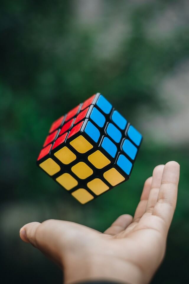
If I asked you to describe to me what you see, you’ll tell me it’s the picture of a hand throwing a cube. How do you know it’s a cube? That’s an assumption. How do you know the hand is throwing the cube? That’s perception. To some, it may look like the hand is about to catch the cube, to some, the hand is throwing the cube. That, my friends, is the difference in what something actually is vs what we perceive. The reason we need to address this concept is because it is fundamental in understanding radiography.
One of the biggest parts of any examination, diagnosis, and treatment planning in Dentistry is the use of dental radiographs. Most, if not all, of our work is based on theoretical knowledge of anatomical structures. We can’t always cut open the tooth or gums to visualize what we are doing and that makes things quite difficult (what an understatement 🙄). Say, to perform a root canal treatment, we assume that we’ll find so and so number of canals at so and so point, to administer an inferior alveolar nerve block, we go by the theory of where the canal ideally would be (must pay attention during anatomy lectures, you guys). With the help of radiographs, we can get a better idea of where and what to find because it is critical to accumulate as much information as possible before commencing irreversible treatment. At the same time, a large amount of our work still relies on blind visualization, mad skills, and tactile sensation along with a few Hail Mary’s.
The hardest bit is to convince patients of the need for radiographs. Every now and then, you come across someone who questions the need for dental x-rays. I recently had a chest x-ray, is this still safe?, My GP wants me to get a CT scan done next week, can we skip this?, My husband and I are planning for a baby, I'd like to avoid x-rays, please. Let’s address those concerns.
So now, what exactly are x-rays?
X-rays, as discovered by Roentgen in 1895, are a high form of electromagnetic radiation (basically a form of energy)1. Sunlight also falls in this category, but visible light only makes up a small portion of the spectrum 1. They are described as consisting of wave packets of energy. Each packet is called a photon, equivalent to one quantum of energy 2. An x-ray is a beam of energy that has the power to penetrate most substances and record image shadows (how cool is that!) on a photographic film or a digital sensor 3.
In a sense, we can say that a radiograph is a two-dimensional representation of a three-dimensional object. When we inform a patient that we’re going to have to ‘take an x-ray’, we basically want to subject them to x-rays to obtain a radiograph. Taking an x-ray is layman terminology for obtaining a dental radiograph.
Why do we need them:
- to aid in detection of lesions and surrounding structures,
- to confirm the presence of a lesion/ disease,
- to provide information which is relevant to treatment planning
- to help evaluate growth and development
- to help identify any changes or variations from what is expected
- to document the condition of a patient at a specific point in time 3
Dental radiographs, as we all know, are of 2 types, mainly - Intra-oral (bitewings, occlusal, periapical) and Extra-oral (OPG, CBCT, lateral cephalometric).
The issue we oft come across is patients refusing radiographs due to fear of excessive exposure to radiation. This situation can be remedied with the following explanation – you get exposed to much higher levels of radiation from a walk in the park on a sunny day than from a dental x-ray (didn’t think about that, did you?).
How’s that, you ask? Let’s talk a little about background radiation.
Let’s face it, radioactivity is a part and parcel of life. To avoid radiation is like avoiding oxygen, it is always present and surrounding us. Natural background radiation has a variety of sources, terrestrial (materials existing in rocks, soil, air and matter), cosmic (from the sun and stars, atmospheric conditions, etc.), internal (radioactive potassium-40 and carbon-14 present in our bodies 4). A huge portion of our exposure is due to radon, which is a gas emanating from the earth’s crust 5.
WHO (World Health Organization) has stated that on average, background radiation constitutes approximately 80% of the annual radiation dose that a person receives 1.
In addition to natural radiation, we are also exposed to human-made sources, aka ionising radiation. Now, this is something we understand and yet, we only actively try and avoid the one most obvious source, which is dental and medical x-rays (fun fact – wearing sunscreen every day, even indoors, can prevent premature aging!).
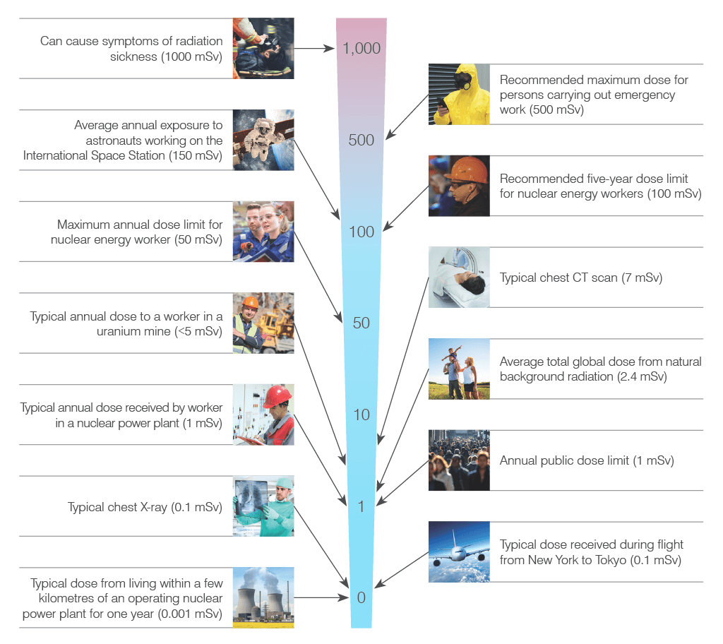
What would also be helpful to know is how much radiation is too much radiation? The SI unit to measure equivalent dose (i.e., the correlation between absorbed dose to effective biological damage) is Sieverts (Sv) 6. The following image 7 is an accurate representation of how insignificant dental x-rays are in terms of contributing to a person’s annual radiation dose.
Whereas, if we had to weight up the amount of radiation exposure from a few common types of radiographs in terms of background radiation, take a look at the following table 8:
| Type of x-ray | Effective dose in mSv (milli-sieverts) | Dose equivalent to days of background radiation |
|---|---|---|
| 1 digital peri-apical radiograph | 0.006 mSv | 1 day |
| 4 dental bite-wing radiographs, F-speed film | 0.038 mSv | 5 days |
| Full mouth x-rays, PSP or F-speed film | 0.171 mSv | 21 days |
| Orthopantomogram | 0.014-0.024 mSv | 2-3 days |
| CBCT (multiple fields of view 9) | 0.003-1.073 mSv | 8-134 days |
| Chest x-ray | 0.170 mSv | 25 days |
| Mammogram | 0.700 mSv | 106 days |
| Head CT (Medical | 2.0 mSv | 243 days |
A brief comparison of the image vs the table helps us deduce that a dental x-ray exposes a person to lesser radiation than travelling in an aero plane (how insane is that!).
Being medical professionals, we are bound by strict codes to comparison provide the most optimal treatment to our patients. To do that, we need the right diagnosis, and consequently the right examination tools. It is very easy to disregard an idea if you don’t know the consequences. Which is why it is of utmost importance to clarify the reasons for exposing a patient to x-rays. The simpler the explanation, the higher the compliance.
P.S. Get them to think about how much radiation they’re exposed to thanks to the blue light emitting from phone screens and laptops 10. Really now, we’re barely exposing them to something they are anyway exposing themselves to.
https://www.who.int/news-room/fact-sheets/detail/ionizing-radiation-and-health-effects ↩︎ ↩︎ ↩︎
https://www.google.co.nz/books/edition/Essentials_of_Dental_Radiography_and_Rad/XO_LDwAAQBAJ?hl=en&gbpv=1&dq=what+are+dental+x+rays&printsec=frontcover ↩︎
https://www.google.co.nz/books/edition/Dental_Radiography_E_Book/w8vsAwAAQBAJ?hl=en&gbpv=1&dq=what+are+dental+radiographs&printsec=frontcover ↩︎ ↩︎
https://www.nrc.gov/about-nrc/radiation/around-us/sources/nat-bg-sources.html ↩︎
https://doh.wa.gov/sites/default/files/legacy/Documents/Pubs//320-063_bkvsman_fs.pdf ↩︎
https://www.arpansa.gov.au/understanding-radiation/what-is-radiation/radiation/measurement ↩︎
https://www.world-nuclear.org/getmedia/280bdda7-182d-49d1-9e96-67f5dc66e2e5/pocketguide_radiation.pdf.aspx ↩︎
https://www.aae.org/specialty/wp-content/uploads/sites/2/2017/07/ecfe-summer-11-final.pdf ↩︎
https://www.orasurgery.com/comparison-of-medical-dental-and-natural-radiation-levels/#:~:text=A%20single%20dental%20x%2Dray,about%2010%20days%20of%20radiation ↩︎